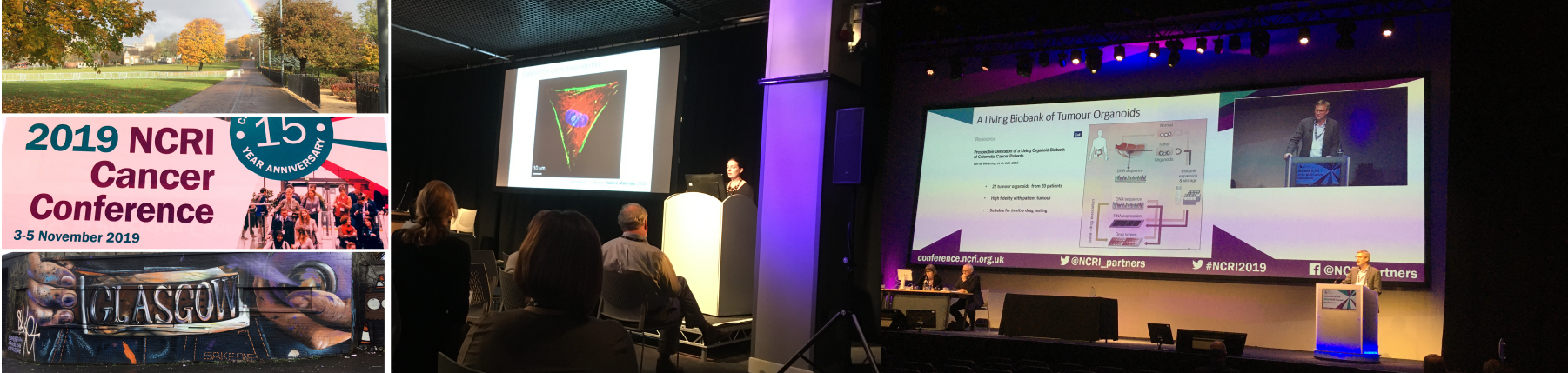
This month the National
Cancer Research Institute (NCRI) conference took place in Glasgow.
Attracting around 1200 attendees, the conference aims to discuss the latest
trends in cancer research and to encourage scientific collaboration worldwide.
Ximbio attended to keep up-to-date with emerging technologies and to identify
new research tools that may benefit from being deposited with Ximbio. Hosted
across three days at the Scottish Event Campus, the programme included a
diverse range of topics. Our session highlights included:
3D models to better study cancer
Investigating the link between obesity and cancer
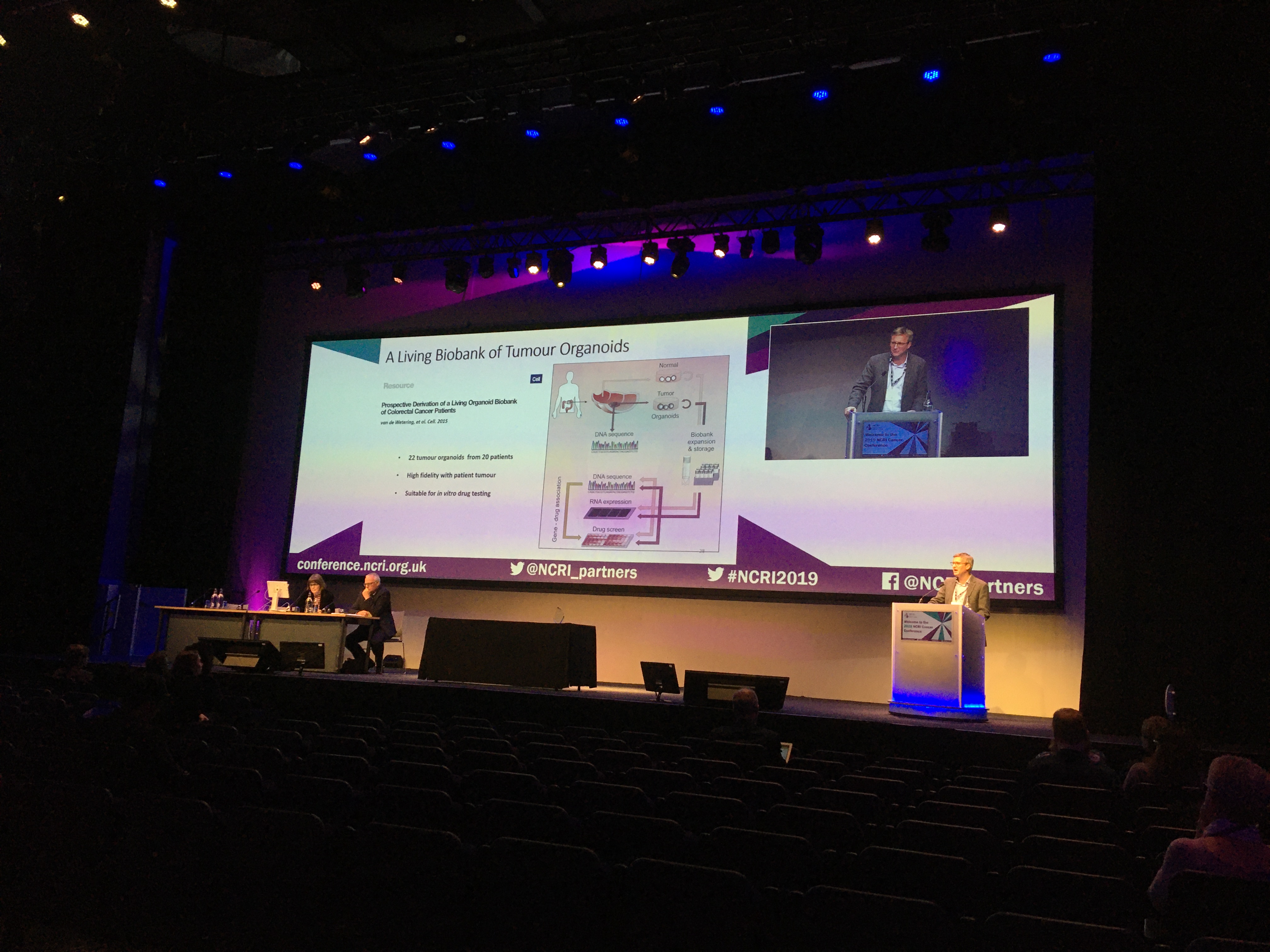
Focusing on: Organoids
It was good to see a project all about increasing the availability of research tools, the Human Cell Model Initiative (HCMI), win the NCRI innovation award. This programme aims to develop a bank of organoid cultures for research use from patient cancer samples. The consortium partners include Cancer Research UK, the Wellcome Trust Sanger Institute (WTSI), the US National Institute of Health (NIH) and the American Type Culture Collection (ATCC). When accepting the award, Matthew Garnett, Translational Cancer Genomics Group Leader at WTSI, described the results of more than three and a half years of research: Over 450 tumour samples have been processed with 130 tumour organoids produced from colon, pancreas and oesophageal cancers and at present 10 tumour organoids from this project are now available from ATCC to order.
How do you make the best cancer cell killers?
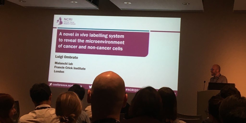
Cancer and its surrounding environment
The environmental context of cancer is a significant area of research interest i.e. the identity of, and how, the non-cancer cells that infiltrate and surround cancer cells may promote or impede proliferation of the cancer cells. This is why a novel metastatic niche-labelling plasmid, presented at the NCRI conference by Luigi Ombrato, from Ilaria Malanchi’s research group, based at the Francis Crick Institute, has attracted much attention. Cell lines stably transfected with the plasmid release a cell-penetrating fluorescent protein which is taken up by neighbouring cells. This enables spatial identification of the local metastatic cellular environment. This plasmid is proving very popular and we have been inundated with requests for it. For more information on this plasmid, visit our website.
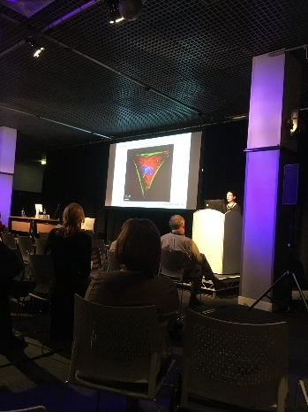
The shape of things to come: Emerging Technologies
A session on emerging technologies for the study of cancer biology, included a presentation by Molly Stevens, Professor of Biomedical Materials and Regenerative Medicine at Imperial College, who described how cell shape and geometry can act as regulators for cell physiology, stem cell signalling and fate. By printing a cell substrate in a triangular shape, a stem cell will adopt the same shape, have greater stiffness and be more prone to differentiate into bone in comparison to a cell in a circular shape.
Hagan Bayley from the University of Oxford presented on emerging technologies for cancer research and showcased a 3D tissue printing technology with the goal to fabricate microtumours for the evaluation of precision therapies; work which has received funding through a Cancer Research UK Pioneer Award. These microtumours could be generated from individual patient’s cells and be used to examine their potential response to therapies. This would help determine what treatment is likely to be most effective for an individual patient.
The 3D tissue printing process involves advanced microfluidics, with each cell inside its own oil drop. These arranged droplets then respond to external stimuli to bring about defined responses/effects. Only a few thousand cells are required and uniform microtumours with more than one cell type can be established. The ultimate aim from this technology is to 3D print tumour cells with customised microenvironments, which could serve as alternatives to organoid/spheroid cancer models.
With such a variety of new topics and trends taking place in
the cancer research environment we look forward to seeing how all this research
progresses over the next year. In the meantime, to find out how we can support
you in your cancer research, why not get
in touch?